
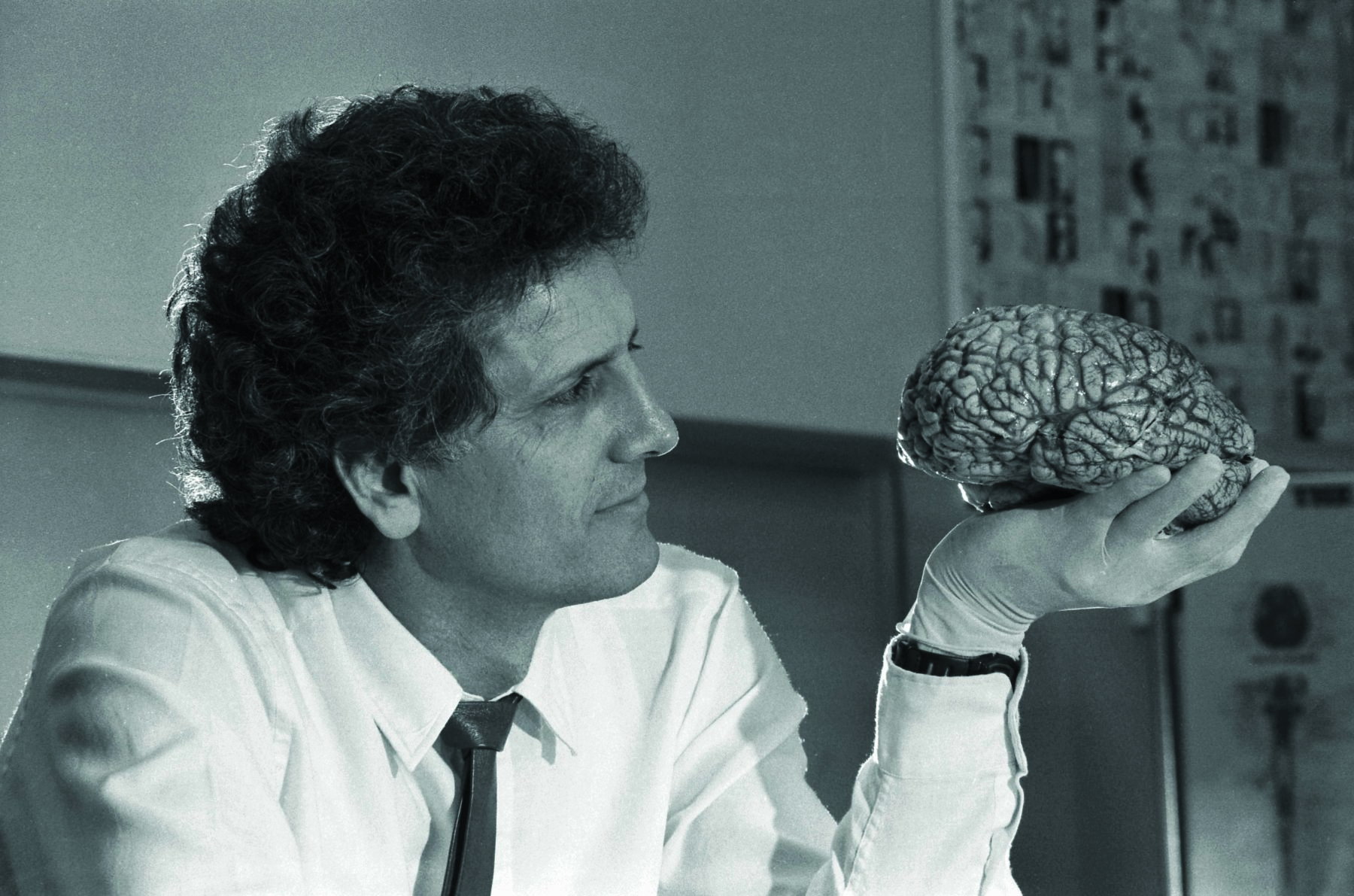
“Never has a bodily organ been so adored as the brain – not since Narcissus looked at his face reflected in water and fell in love with it,” Professor George Paxinos says. “And never was there so little justification for it.” It’s a surprising comment from a man who’s spent his life mapping every nook and cranny of the brain, studying its every structure and cell type. George, born in Ithaca in Greece, has authored or co-authored 57 scientific books on the organ, including what for three decades was the third most cited scientific book ever published. He’s also identified and named more brain areas than anyone.
George isn’t a fan of what he sees as almost cult worship of the organ, which is made up of some 1.2–1.4kg of pink-grey tissue. But since he discovered that a particular chemical was able to colour brain structures, allowing researchers to produce images that display them like countries on a world map, George has obsessed over the organ’s minutiae, seeking out the borders between those neurological nations with ever increasing precision and detail.
His books and papers are used worldwide as a guide to steer anyone with a professional interest in the organ – from brain surgeons to biology students – through the cellular mazes of mouse, rat, bird, monkey, chimpanzee and human brains and spinal cords. The accolades with which his work has been honoured include an Order of Australia, a Humboldt Research Award and the NSW Premier’s Prize, and he has been elected a corresponding member of the prestigious Academy of Athens – only the second living Australian to achieve the honour.
Despite his extensive and highly accomplished academic career, George himself says his finest achievement is a 384-page novel entitled A River Divided, which took him 21 years to write and was finally published in October 2021.
THE PAXINOS LAB AT Neuroscience Research Australia (NeuRA), in Sydney’s eastern suburbs, is remarkable in its ordinariness, given the impact it’s had on our scientific understanding of the central nervous system. Fluorescent lighting washes over tables piled with papers and books, there are a few computers around the edge of the room, which seems surprisingly small, and the walls are lined with shelves and filing cabinets.
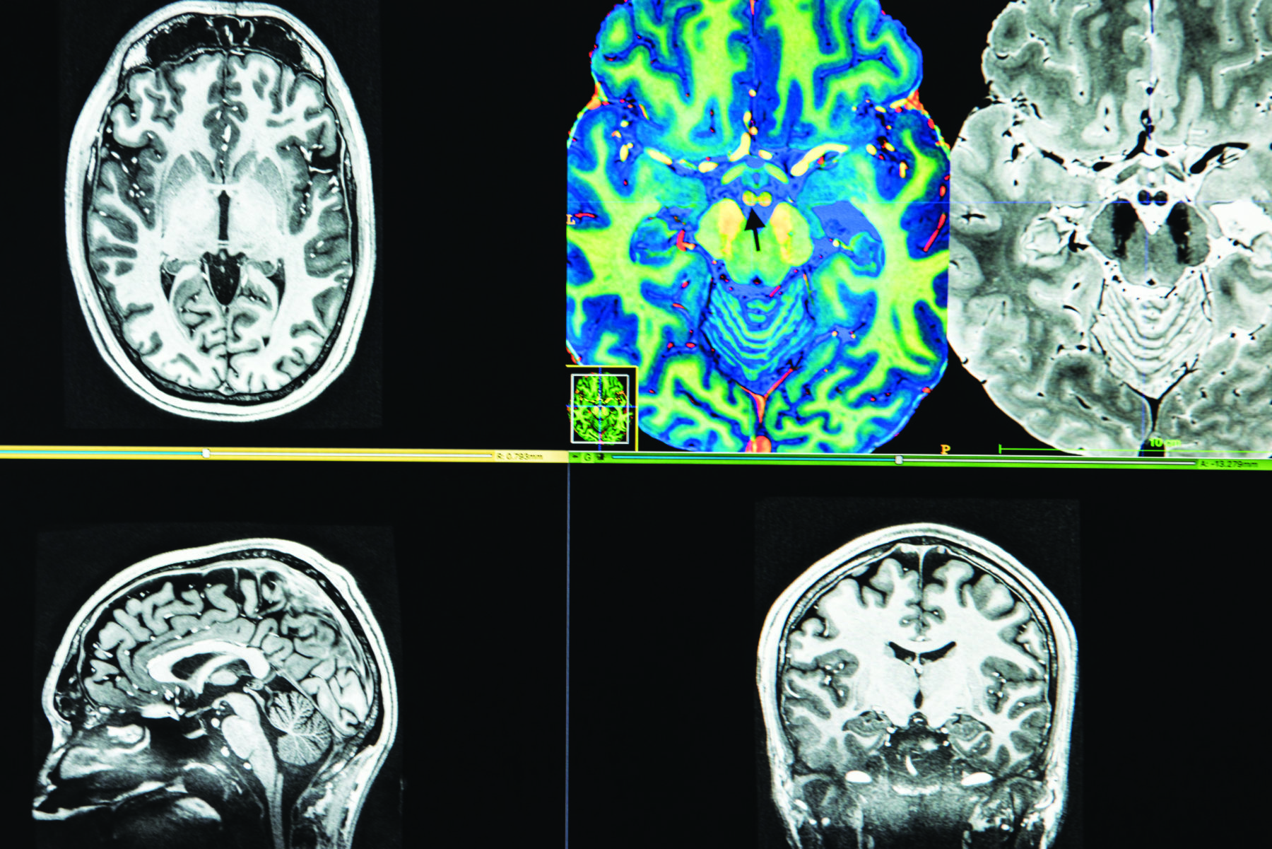
Clues as to what goes on here include: a larger-than-life colourful plastic model of a brain on a shelf, vibrant fluorescent magnetic resonance imaging (MRI) pictures of a brain – belonging to one of George’s colleagues, it turns out – strewn across the table, and if you look closely at the small wooden and plastic boxes on another shelf, the labels read: “HUMAN”, “MONKEY” and “RAT”.
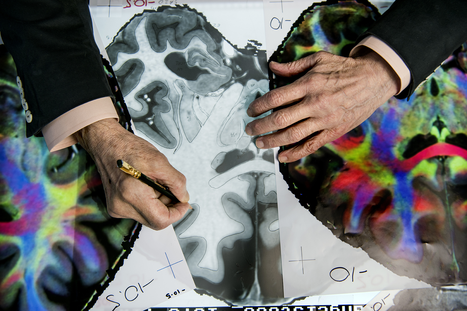
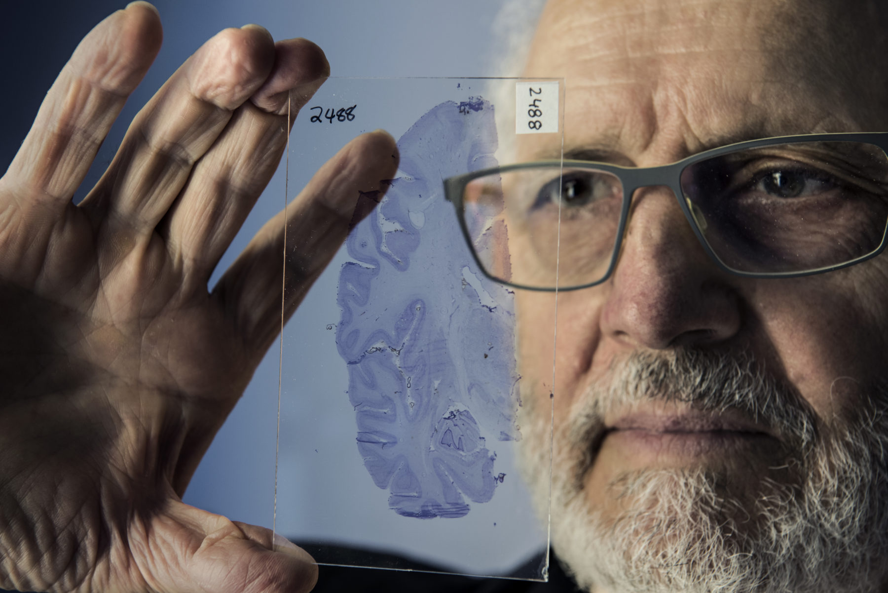
These boxes are full of microscope slides embedded with wafer-thin slices of central nervous system tissue. Between the shelves and the refrigerators in a nearby room, the lab has about 80,000 slides of brain and spinal cord tissue, each coloured with a chemical or stain designed to highlight particular features, ranging from cell types to neurotransmitter concentrations.
A stain like this was what started George on his long journey to scientific fame. While studying mathematics at university in his birthplace, George undertook an elective unit of psychology and was surprised to find he enjoyed it.
I thought, Why shouldn’t I go where my nose is going?“
George says.
His nose took him to the University of California, in the USA, to study psychology, then to McGill University in Montreal, Canada, where he completed a PhD.
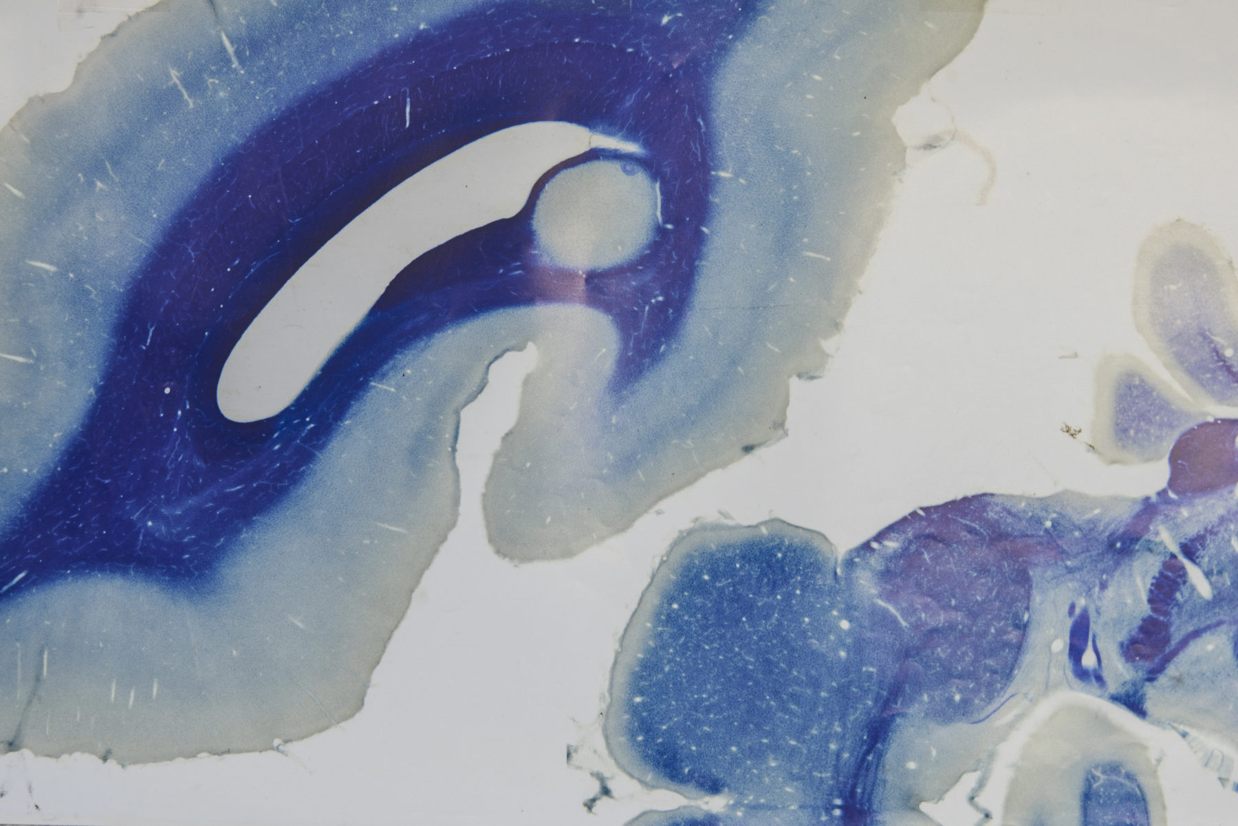
While George was on sabbatical at the University of Cambridge, England, in 1977, he came across a chemical that demonstrated the location of the brain enzyme acetylcholinesterase, which breaks down the neurotransmitter acetylcholine that carries nerve impulses across the junctions between neurons.
This stain revealed the organisation of the brain like a child’s colouring book. “And although I was not an anatomist, I thought I would be able to construct an atlas of the brain of that rat…[that was] better than the existing atlases,” George recalls. His first book, co-authored with long-time research colleague Charles Watson – The Rat Brain in Stereotaxic Coordinates – was published in 1982. Today, it is one of the most cited books of all time and is up to its seventh edition.
From rat to human, via monkey and chimpanzee, is not as big a leap in brain structure as one might imagine. An area that shows up brown with the acetylcholinesterase stain in a particular brain region in the rat can be linked to a similar stain-dense region in a mouse, chimpanzee, and even human. And once one brain region is outlined with this particular stain and its borders established, it can then be mapped in relation to other known regions of the brain.
George started with the acetylcholinesterase stain, but one of his more recent books uses eight different stains, each of which highlights different aspects of the brain and reveals ever more fine-grained detail.
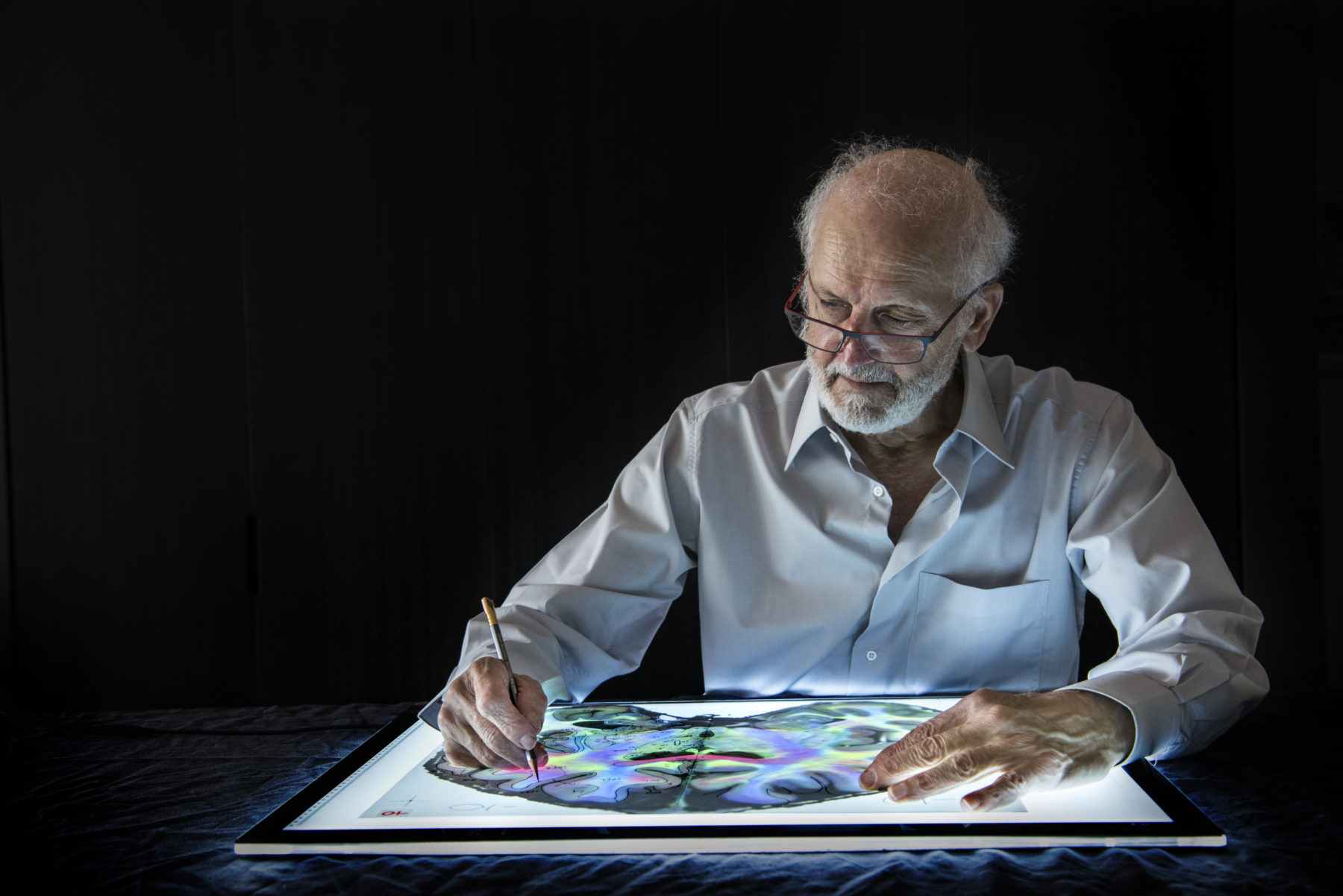
George’s latest project – the brilliantly coloured workings of which are currently scattered across surfaces all over the room – is a book that uses MRI pictures to show the living human brain in the highest resolution yet. The fluoro colours reveal which neurons in the image span from top to bottom, which span from side to side, and which span from front to back. It will enable detailing of connections between different parts of the brain.
It’s the first in-depth anatomical project using a living brain – two of them, in fact. One belongs to postdoctoral fellow Dr Steve Kassem, who puts up with good-natured teasing about the fact that the MRI scanner had to be adapted for his large head. Steve spent 40-plus hours having his brain imaged; those hours have generated hundreds of pictures, each a fine electronic slice through the human brain.
George and his colleagues take a surprisingly low-tech approach to analysing and assembling their atlases: they lay the images out sequentially, beginning in one corner of the office. They then spread them out across the desk then along the floor, out into the corridor and into the next room so that they can “walk” through the brain and see where different structures start and end. George points to one trail of images. “You can see this structure commences here,” he says. “We keep going, it gets bigger, bigger, then smaller and smaller and disappears.”
Another low-tech method involves tracing paper. Steve explains they trace the initial lines around each section revealed by the stain or scanner, but then they can lay the outlines on each piece of paper over images created using different stains to reveal the connections and overlaps. Just because it’s low-tech doesn’t mean it’s straightforward; the tracing paper has to be imported because it’s no longer made in Australia.
It seems an obvious opportunity to assemble images into a three-dimensional electronic model using the wealth of imaging software, programs and even artificial intelligence algorithms now available to scientists working in this and many other fields. But research assistant Dr Kristie Smith says these technologies wouldn’t be equal to George at displaying the finer detail.
“It’s the technique George uses that lets you get into the nitty-gritty,” Kristie says.
It’s because of work like this that you can pick up what machines do miss. Until we have a machine that’s as intelligent as us, we will still have a job,”
George says.
This poses the question of whether these maps are really just the brain according to George Paxinos. Steve is quick to point out that George’s work has been cited by other scientific papers more than 100,000 times, “which is considered a benchmark for being one of the most prominent scientists in the world”, he says. George, who’s received only one piece of fanmail during his illustrious career, knows true praise for his work lies in his record of citations. “If they cite your work, that means they agreed with you, otherwise they wouldn’t use you,” he says.
George’s lab is not the only neuroscience group working on mapping the brain. It has stiff competition from some scientific heavy hitters, notably the Allen Institute for Brain Science, the scientific division of Seattle’s Allen Institute funded by the late Paul Allen, co-founder of Microsoft. An independent study that compared the two research groups’ brain-mapping efforts on the mouse brain concluded that George’s maps were more accurate. And that was using an earlier edition of his book – the third edition, George interjects with quiet pride – which was more than a decade old by the time. “A $200 million institute still can’t compete against him,” Steve says.
WHY DOES AN eminent brain cartographer with 57 scientific books to his name spend 21 years writing a science fiction novel? George says he has had just two goals in life: to produce the atlas of the rat brain, and to write this novel. But the motivation came not from his scientific pursuits – although the book is steeped in neuroscience and genetics – but from his environmental conscience.
On his own website George describes himself as an environmental activist. He has a history of environmental campaigning; from 1989 until 2000, he was a key advocate for the return of trams to Sydney, and he founded the Light Rail Association, which aimed to reduce atmospheric pollution caused by petrol-powered vehicles. He organised demonstrations and fought hard to preserve Sydney’s tramway infrastructure – its sidings, workshops, corridors and even the trees – for the future light rail. But despite working for hours on the campaign each week, he failed. “I thought that rather than working to stop someone from cutting a tree by placing barriers around it, if I managed to work upstream to change their attitude to make them not want to cut the tree, then that would be more effective,” he says. He decided writing a novel was the way to do it, but at the time he didn’t know what to write about.
The idea came to him at a Christmas party in 1999. George was about to travel to Spain, and someone suggested he could visit the bones of Saint James, which are housed in the Cathedral of Santiago de Compostela. His first thought was that it must be possible to extract some DNA from those bones and see what the man looked like. “And then I thought, Why not someone far more important?”
That someone was Jesus of Nazareth. The premise of the novel is that a geneticist and amateur archaeologist discovers the remains of Jesus in the desert near Masada, in Israel, secretly extracts DNA and creates two embryos. The resulting babies are raised by surrogate mothers – one in wealthy surroundings in Sydney, and the other in the slums of Santiago, Chile. “It asks the question, ‘What if Jesus was here today – who would he side with? With BHP? With Adani? Or with the guys who tried to stop it?’” George says.
The novel resonates with George’s environmental passions, but also with his beliefs about the idea of free will. When we talk about the brain as being the seat of the individual, he is quite clear that he does not hold the brain in any kind of awe, as a repository for the soul and all that makes us who we are.
“The brain makes decisions, but the problem is that the decisions it makes are based on two and only two things: genetic predisposition from the genetic endowment we receive and the environmental influences that have shaped that endowment,” George says. “So there can be no freedom: the brain will make the decision through its physics – that is, neurotransmitters, receptors, enzymes and electrical activity – and this decision will be based exclusively on the experiences and genetic endowment up to this point.”
While not holding the brain in higher esteem than any other part of the body, George does recognise it to be the source of what makes us ‘us’. “If the soul is supposed to be where stimulation becomes perception, where reasoning takes place, where decisions are made, where love is manufactured, where memories are stored, then there is no reason to postulate its existence, because there is already an organ that does these things,” he says. “It’s called the brain.”


